
Biological samples require a hydrated environment to maintain their structure and retain dynamic behavior. With Protochips’ machine-vision powered suite of in-situ TEM tools you can utilize the electron microscope as a real-time laboratory to study delicate, low contrast systems at a resolution of a few nanometers.
Poseidon AX: Liquid, electrochemical and liquid heating in situ studies
Study nanoscale dynamics, interactions, and processes of virus, cells, proteins, micelles, and other biological and biomimetic materials while maintaining a physiologically relevant environment.
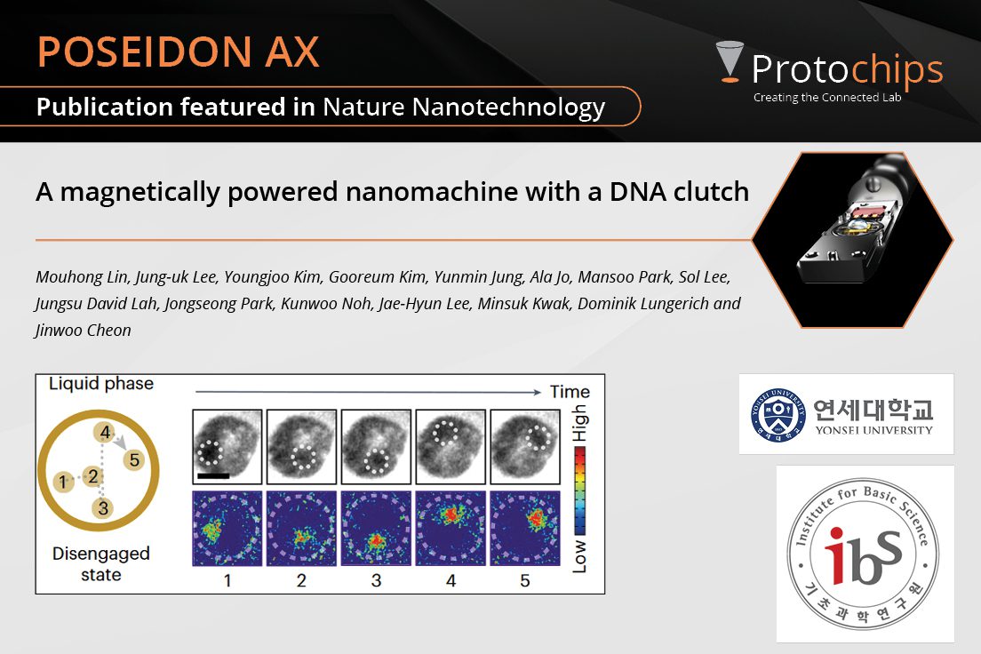
Lin, M. et al. (2024) 19, 646–651

Featured Papers
-
News
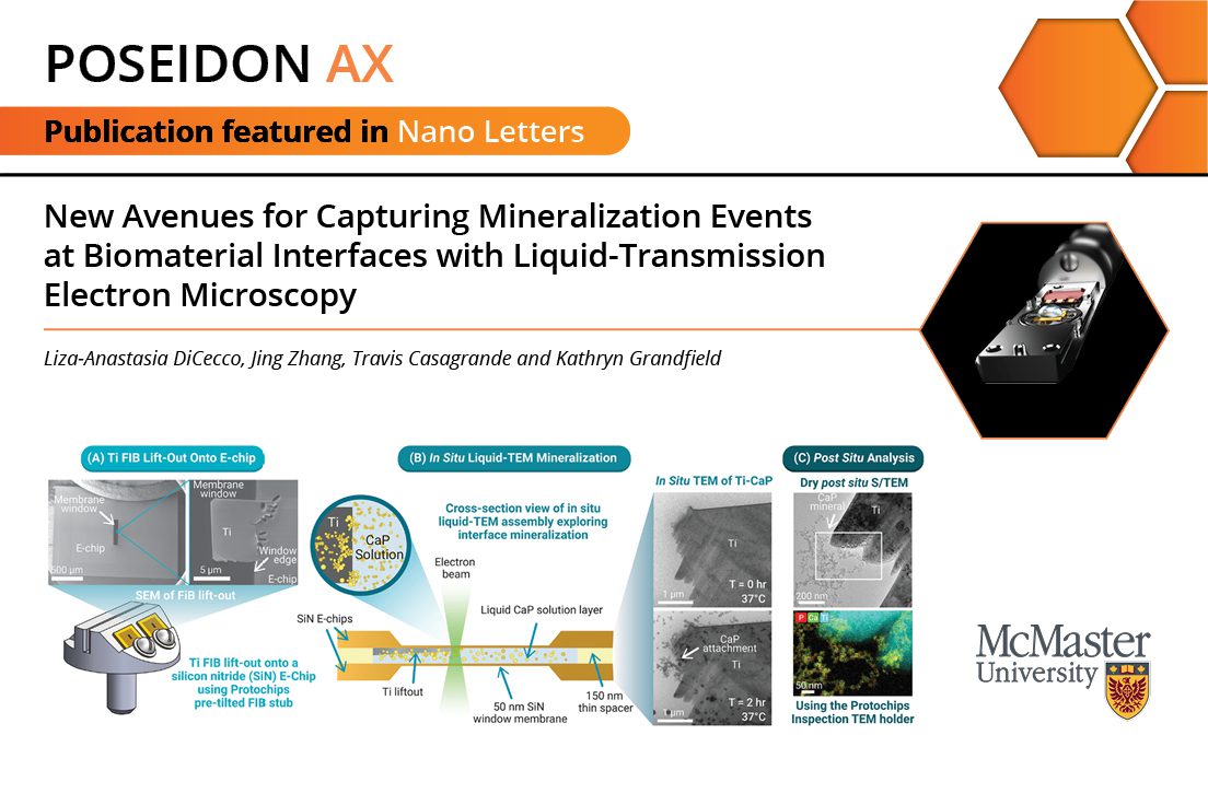
New Avenues for Capturing...Oct. 22, 2024 -
News
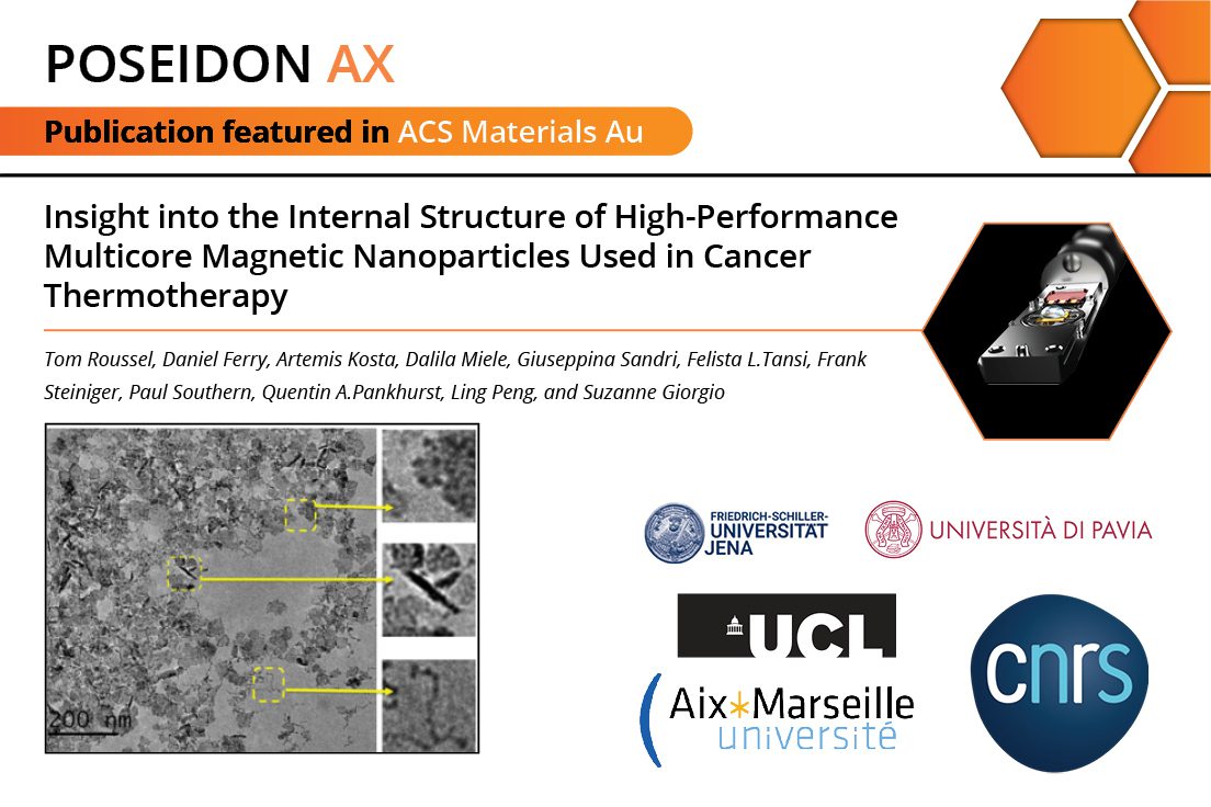
Insight into the Internal...Sep. 10, 2024 -
News
A magnetically powered na...Apr. 9, 2024 -
News
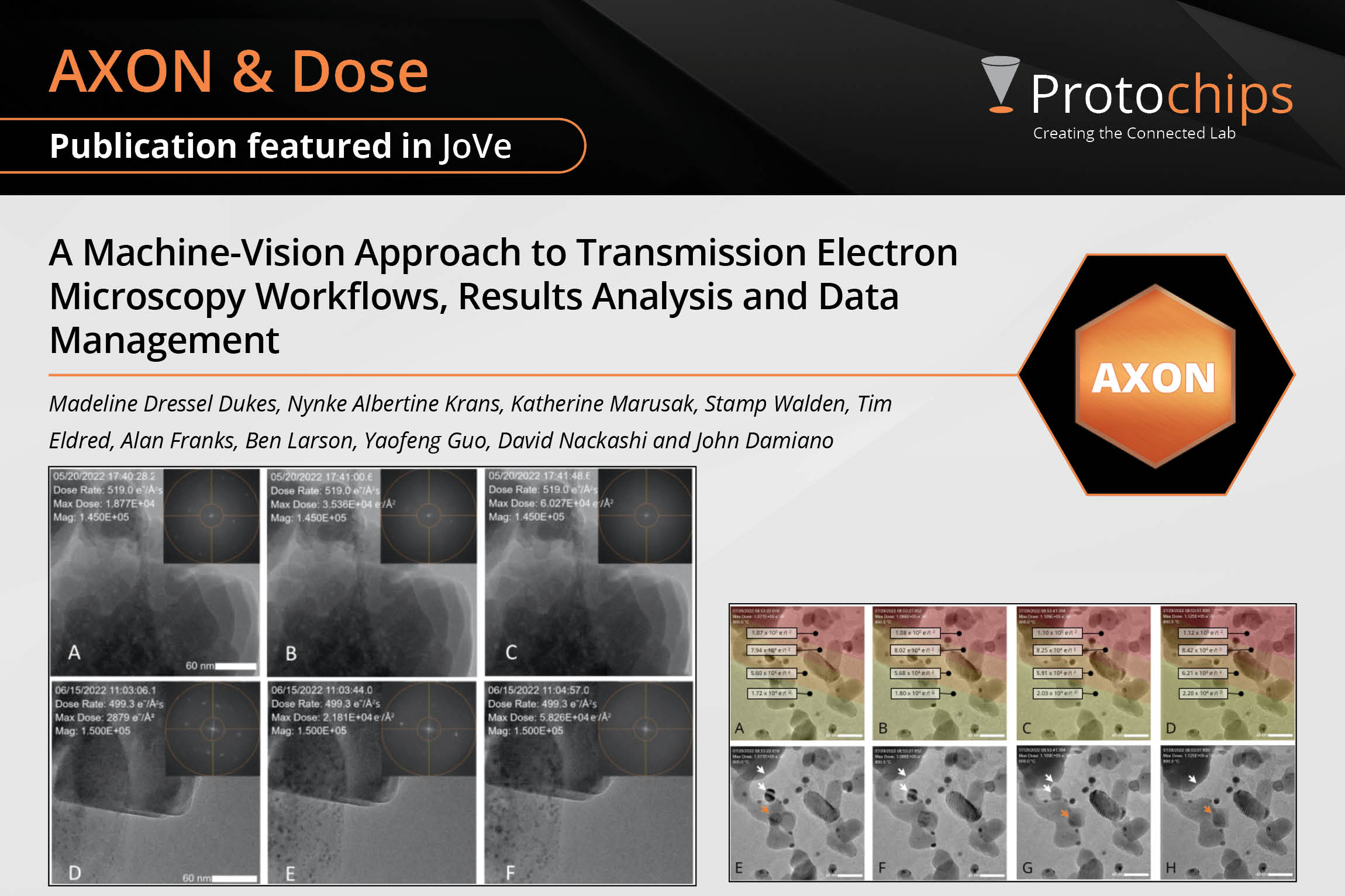
A Machine-Vision Approach...Sep. 5, 2023 -
News
Degratation Mechanisms of...Aug. 29, 2023 -
News
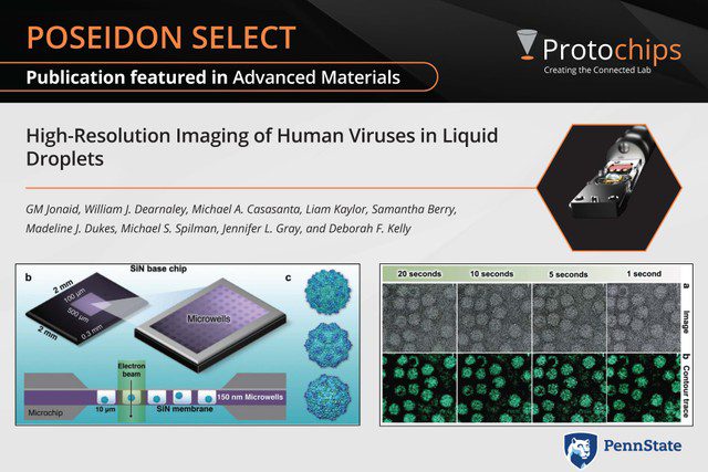
High-Resolution Imaging o...Dec. 13, 2022 -
News
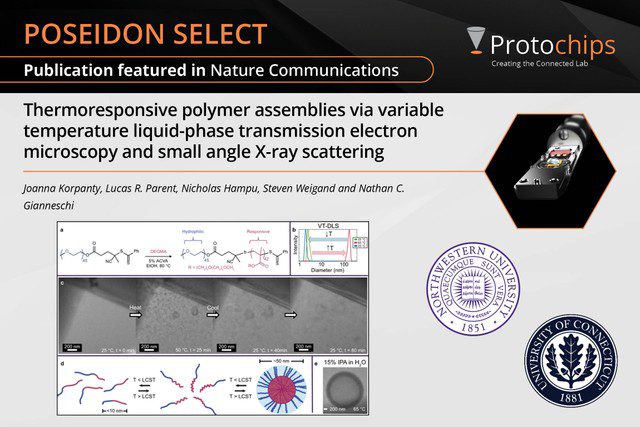
Thermoresponsive polymer ...Dec. 6, 2022 -
News
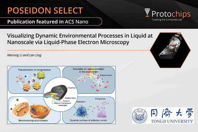
Visualizing Dynamic Envir...Nov. 18, 2022 -
News
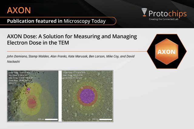
A Solution for Measuring ...Nov. 15, 2022 -
News
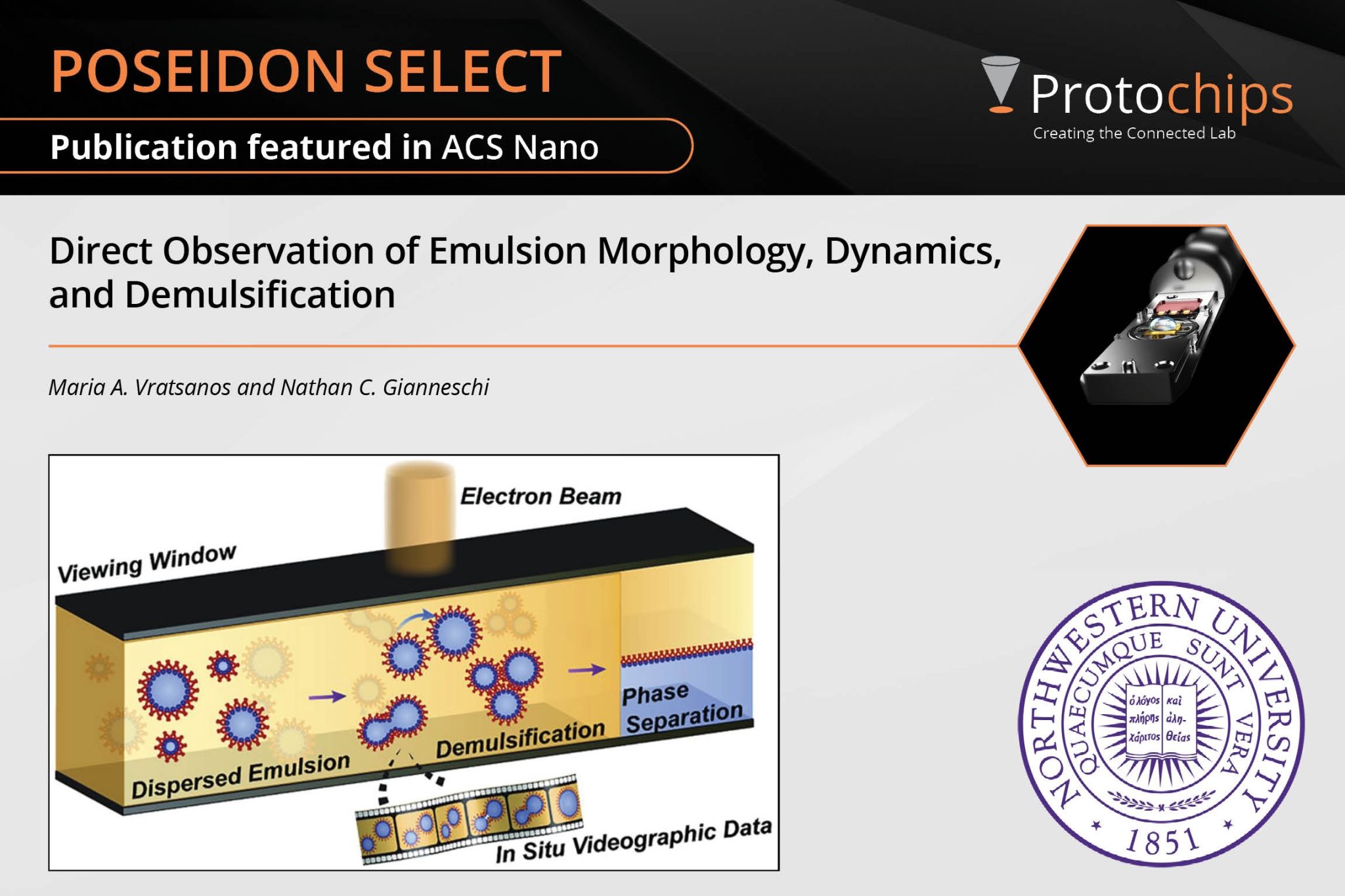
Direct Observation of Emu...Oct. 17, 2022
What have our users done in the field? Read our summaries here!
-

Biological Materials One Pager
Download the one pager that shows how Poseidon AX can be used to study biological materials with in situ electron microscopy.
-
Radiolysis & Liquid-EM
In this application note, written by Protochips, we summarize how in situ electron microscopy using liquid phase is challenged by radiolysis side effects and how to minimize these effects.
-
Using Protochips AXON Software for Tracking Electron Flux and Cumulative Dose
In this paper, we take a look at how AXON Dose tracks the electron flux and cumulative dose, and why this might be useful for all experiments.
-
Scanning Transmission Electron Microscopy of Live Yeast Cells in Liquid
In this summarized paper, Dr. Diana B. Peckys was able to image live yeast cells in hydrated form in the electron microscope using the Poseidon system.
-
Correlative Light and Electron Microscopy (CLEM) of Eukaryotic Cells in Liquid
In this summarized paper, researchers from the group of Dr. Niels de Jonge cultivated COS -7 fibroblast cells directly on Poseidon E-chips.
-
Understanding the Mechanism of Carbon Nanotube Degradation in Macrophage Cells
In this summarized paper, researchers from the group of Dr. Alloyeau looked at the stability of carbon nanotubes in different liquids to understand the degradation of these materials in macrophage cells.
Videos
Watch dynamic behavior of real samples in situ.
This movie shows a series of images that demonstrates the tilt range of Poseidon Select. Images of aggregated 30 nm gold nanoparticles in 1.5 μm of water were collected in 5° increments, over a total angle of -30° to +30°. Data was collected using a Philips/FEI CM300FEG TEM equipped with a GIF energy filter at 300 kV.
Electron beam-induced growth of calcite nanoparticles in situ in the presence of AP7, a nacre protein that is found in abalone shellfish. The calcite particles were grown in the spatially confined conditions of the in-situ liquid cell in a liquid layer of 500 nm. During growth both a stable protein mediated scaffold-type structure, and an unstable crystalline structure which re-dissolved shortly after formation, were observed. (JEOL JEM-2200 FS with double Cs correction, operated at 200 KV in STEM mode. Movie playback is increased by a factor of 25). Courtesy of Andreas Verch and Roland Kroeger. For more information on Poseidon Select, go to our website at http://www.protochips.com/products/poseidon.html
AXON Synchronicity enables live drift correction of vesicular structures in fabric softener. Data was acquired using a Gatan 626 cryogenic holder on a Tecnai G2 Twin at the Shared Materials Instrumentation Facility at Duke University. This movie demonstrates the power of AXON’s image stabilization even at cryogenic temperatures with a non-Protochips holder. For more information about AXON Synchronicity, visit us at https://www.protochips.com/solutions/tem-data-correlation/tem-software/.
Additional information: movie was roughly 10min and acceleration voltage was 200kV
Learn about Protochips' groundbreaking liquid cell for in situ TEM and STEM.
#FindYourBreakthrough | FLASH TALKS: EP #4
In situ monitoring of exopolymer-dependent Mn mineralization on bacterial surfaces
Presented by: Dr. Thaïs Couasnon and Dr. Damien Alloyeau from Université de Paris, CNRS. Read the full paper here: https://advances.sciencemag.org/content/6/27/eaaz3125#BIBL
For more product information, visit our website at www.protochips.com or reach out to us at https://www.protochips.com/contact-us/
#FindYourBreakthrough | FLASH TALKS: EP #11
Monitoring of CaCO3 Nanoscale Structuration through Real-Time Liquid Phase Transmission Electron Microscopy and Hyperpolarized NMR
Presented by: Dr. Vinavadini Ramnarain from IPCMS/University of Strasbourg. Read the full publication here: https://pubs.acs.org/doi/10.1021/jacs.2c05731?ref=pdf
This study focuses on advanced magnetic nanoparticles (MNPs), made of iron oxide cores embedded in sugar or starch matrices, with remarkable heating capabilities crucial for clinical applications like pancreatic cancer treatment. Using cutting-edge electron microscopy techniques and the PoseidonAX system, Dr. Tom Roussel reveals insights into the particles' structure: well-dispersed iron oxide cores and unique dextran lamellae arrangements. Magnetic analysis linked these structural features to the particles' exceptional heating performance. This research highlights how precise structural design can optimize MNPs for future cancer therapies.
Read the full paper here!
https://pubs.acs.org/doi/10.1021/acsmaterialsau.4c00021
Presented by :
Tom Roussel, PhD
Interdisciplinary Center of Nanoscience of Marseille (CINaM)
This flash talk discusses how recent advances in liquid phase scanning transmission electron microscopy (LP-STEM) are overcoming challenges like sample thickness and beam damage to revolutionize live-cell imaging. Using graphene encapsulation and advanced imaging techniques with the AXON Synchronicity software, Dr. Brian Caffrey shows details of cellular ultrastructure, demonstrated with the resilient bacterium Deinococcus radiodurans. Watch this flash talk to learn how LP-STEM is opening new frontiers in biological research!
Want to read the entire work?
Find it here!
https://onlinelibrary.wiley.com/doi/10.1002/smll.202402871
Presented by :
Brian Caffrey, PhD
The Rosalind Franklin Institute